SUMMARY
Early Events in Tooth Development
Vestibular and Dental Lamina
- At about the 6 weeks, basal cells of the oral ectoderm proliferate rapidly, forming the Primary epithelial band.
- At the 7th week, the epithelial band divides into:
- Dental lamina (lingual side)
- Vestibular lamina (buccal side)
Dental Lamina
- It is the lingual process of the Primary Epithelial Band.
- Acts as the primordium of the ectodermal portion of deciduous teeth.
- Permanent molars arise from a distal extension of the dental lamina.
- Successors of deciduous teeth arise from the lingual extension of the free end of the dental lamina (called Successional Lamina).
- The lingual extension develops from the 5th month in utero (central incisor) to the 10th month of age (second premolar).
- Total activity of dental lamina extends for at least 5 years.
Vestibular Lamina
- Located labial and buccal to the dental lamina in each dental arch.
- Also known as the Lip Furrow Band.
- It proliferates buccally → enlarges → undergoes cell apoptosis → forms the oral vestibule.

Oral Cavity and Development of Dental Lamina
- The primitive oral cavity, or stomodeum, is lined by stratified squamous epithelium called the oral ectoderm or primitive oral epithelium.
- The oral ectoderm contacts the endoderm of the foregut to form the buccopharyngeal membrane. At about the 27th day of gestation, this membrane ruptures.
- Two or three weeks after rupture of the buccopharyngeal membrane, when the embryo is about 6 weeks old, certain basal cells of the oral ectoderm proliferate rapidly and form the primary epithelial band.
- At about the 7th week, the primary epithelial band divides into an inner (lingual) process called dental lamina and an outer (buccal) process called vestibular lamina.
- Dental lamina serves as the primordium for the ectodermal portion of the deciduous teeth.
Fate of Dental Lamina
- It extends over a period of at least 5 years.
- It may still be active in the molar region.
- Remnants of dental lamina persist as epithelial pearls or islands within the jaw as well as in the gingiva, referred to as CELL OF SERRES (sometimes it may proliferate and lead to odontogenic cyst & tumors).


Tooth Formation Timeline
- Expression of Lhx genes is the earliest molecular event.
- Lhx gene (Lhx-6 and Lhx-7) expression occurs at the 9th day of intrauterine life.
- 1st genetic evidence (molecular evidence) of tooth formation ⇒ 9th day of intrauterine life.
- Initiation of epithelial band formation ⇒ 11th day of intrauterine life.
- 1st histological evidence of tooth formation ⇒ 11th day of intrauterine life.
- Completion of epithelial band (37th–39th day) ⇒ 6th week IU.
- 1st evidence of tooth formation ⇒ 6th week IU.
- Proliferation of dental lamina occurs from 7th week (i.e. 48th–49th day IU).
- After 7th week IU, vestibular lamina forms.
- By 11th week – Tooth bud becomes visible to the unaided eye.
- 1st macroscopic evidence of tooth formation ⇒ 11th week IU.
- At 14th week ⇒ Initiation of calcification occurs.
- 1st radiographic evidence of tooth formation ⇒ 14th week IU.
Tooth Development Stages
Overview
Tooth development is a sequential process that includes both physiological and morphological stages. These stages are essential to form the proper structure, shape, and functionality of teeth.
Physiological Stages
- Initiation
- Proliferation
- Histodifferentiation
- Morphodifferentiation
- Apposition
Morphological Stages
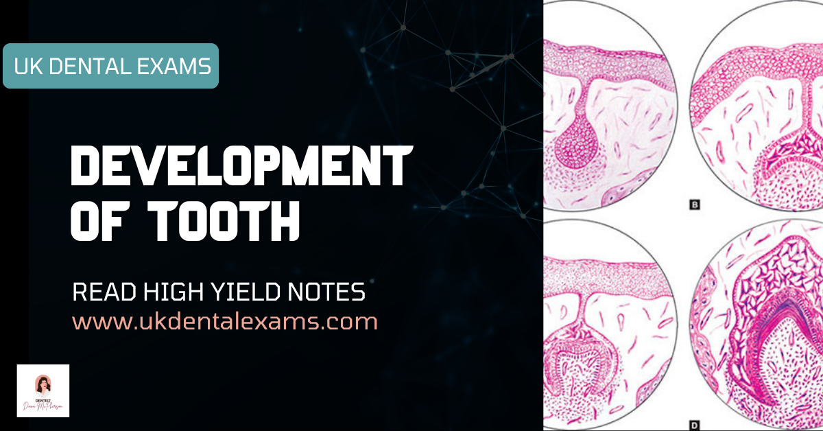
- Dental Lamina
- Bud Stage
- Cap Stage
- Bell Stage
- Early Bell Stage
- Advanced Bell Stage
Bud Stage
This is the first morphological stage. The dental lamina gives rise to the tooth bud. The bud consists of peripheral low columnar cells and central polygonal cells. At this stage, the dental papilla and dental sac begin to form.
Cap Stage
In the cap stage, three distinct layers develop: the outer enamel epithelium (OEE), stellate reticulum, and inner enamel epithelium (IEE). The cap-shaped enamel organ surrounds the dental papilla and sac. Cells differentiate into low cuboidal and star-shaped forms, contributing to the early enamel structure. Key structures formed in this stage include the enamel knot, enamel cord, and enamel septa.
Early Bell Stage
This stage is characterized by increased histodifferentiation and morphodifferentiation. The four layers of the enamel organ become more distinct: OEE, stellate reticulum, stratum intermedium, and IEE.
- Stratum intermedium becomes rich in glycogen, aiding in enamel formation.
- Collapse of stellate reticulum reduces the distance between ameloblasts and OEE capillaries, facilitating nutrient transfer.
- OEE folds inward, allowing the dental sac to condense and form vascular papillae which nourish the developing enamel organ.
Advanced Bell Stage
This stage marks the beginning of mineralization and root formation. The four primary layers remain, but the future dentinoenamel junction (DEJ) is now defined. The cervical portion of the enamel organ develops into the Hertwig’s epithelial root sheath (HERS), which plays a critical role in root formation.
Summary
Tooth development follows a tightly regulated sequence of events from the initial epithelial thickening (dental lamina) to the formation of mature enamel and roots. Each stage is interdependent, and defects in any phase can result in dental anomalies affecting structure, number, or shape of teeth.


Developmental Stages of Tooth
Bud Stage
- Proliferation of dental lamina cells leads to localized swelling in connective tissue → bud shape appears in the dental lamina → this stage is called the Bud Stage.
- Marks initiation of tooth formation.
- No histodifferentiation at this stage.
- Only epithelial cells are present (same morphological cell types).
- Structures such as Dental Papilla, Dental Sac, Inner Enamel Epithelium, Outer Enamel Epithelium, and Stellate Reticulum are absent.


Cap Stage of Tooth Development
Unequal growth in different parts of the tooth bud leads to the cap stage, which is characterized by a shallow invagination on the deep surface of the bud.
→ Marks the beginning of histodifferentiation.
Cell Types in Cap Stage
- Outer enamel epithelium
- Inner enamel epithelium
- Stellate reticulum
Outer Enamel Epithelium
- Peripheral cells are cuboidal, cover the convexity of the cap.
- Separated from the dental sac and papilla by a basement membrane.
Inner Enamel Epithelium
- Cells in the concavity of the cap become tall, columnar.
- Enamel organ has a double attachment of dental lamina to the oral epithelium enclosing ectomesenchyme, called the enamel niche.
Stellate Reticulum
- Polygonal cells begin to separate due to osmotic force exerted by glycosaminoglycans.
- They become star-shaped (Stellate cells).
- Maintain contact via cytoplasmic processes forming a network — Stellate Reticulum.
Transitional Structures
Enamel Knot
- Appears at the end of bud stage → marks transition of bud stage to cap stage.
- Expresses growth factors:
- Slit-1
- FGF-4, 9
- BMP-2, 4, 7
- Acts as a reservoir of dividing cells for the enamel organ.
- Plays a key role in determining shape of the tooth:
- Main component for cusp shape
- Functions as organizational center
Enamel Cord
- A vertical extension of the enamel knot.
- Also acts as a reservoir of dividing cells.
Enamel Septum
- When the enamel cord extends to meet the outer enamel epithelium, it forms the enamel septum.
- This structure divides the stellate reticulum into two parts.

Bell Stage of Tooth Development
Crown shape is determined in this stage.
- Attributed to pressure from growing dental papilla cells on IEE, opposed by stellate reticulum fluid.
- Different mitotic rates cause folding of enamel organ → different crown shapes.
- IEE cells at future cusp tip/incisor region divide first and stop dividing; this progresses cervically (last to differentiate).
- Tooth morphogenesis is under the control of:
- Genes and their signaling molecules
- Growth factors
Inner Enamel Epithelium
- Single layer of cells → differentiate into tall columnar cells called ameloblasts.
- Ameloblast size: 4–5 µm diameter and 40 µm long.
- Organize underlying mesenchymal cells in dental papilla → later become odontoblasts.
Stratum Intermedium
- Located between IEE and stellate reticulum.
- Squamous cells with cytoplasmic organelles, acid mucopolysaccharides, and glycogen deposits → metabolically active.
- Essential for enamel formation.
Stellate Reticulum
- Before enamel formation, stellate reticulum collapses → brings ameloblasts close to outer enamel epithelium for nutrition.
Outer Enamel Epithelium
- Folds outward; adjacent dental sac forms papillae with capillary loops.
- Provides rich nutritional supply for avascular enamel organ.
- Nutrition to ameloblasts is provided by dental sac (later replaced by diffusion through enamel).
Dental Papilla
- Peripheral mesenchymal cells become odontoblasts under epithelial influence.
- Dental papilla gives rise to dentine and pulp.
Dental Sac
- Fibers differentiate to form PDL.
- PDL fibers get embedded into cementum and bone.
- Dental sac gives rise to PDL.


Advanced Bell Stage
- This stage marks the beginning of mineralization and root formation.
- The interface between inner enamel epithelium and odontoblasts establishes the dentinoenamel junction.
- The cervical portion of the enamel organ forms the epithelial root sheath of Hertwig.
- Hertwig’s epithelial root sheath (HERS) guides the development of the root’s shape, length, size, and number.

Stages of Tooth Development
| S. No. | Stages | Layers | Cells | Events |
|---|---|---|---|---|
| 1 | Bud stage | – |
|
|
| 2 | Cap stage |
|
|
|
| 3 | Early bell stage |
|
|
|
| 4 | Advanced bell stage |
|
|
|

Tooth Development: Enamel Organ & Related Structures
- Initiation of primary teeth begins around 4–6 weeks I.U. (first trimester of pregnancy).
- Initiation of permanent teeth and initiation of calcification of primary teeth is in second trimester of pregnancy.
- Enamel organ – forms enamel. It consists of:
- Outer enamel epithelium (OEE) – Provides nutritional supply.
- Inner dental epithelium (IEE) – Gives rise to Ameloblasts which produce enamel.
- Stratum intermedium – Cells show unusually high activity of alkaline phosphatase, essential for amelogenesis. Also demonstrates high level of amino peptidase during dentinogenesis.
Enamel Knot
- The cells in the center of the enamel organ are densely packed and form the enamel knot.
- The enamel knot and cord may act as a reservoir of dividing cells for the growing enamel organ.
- Enamel knot precursor cells can be detected first at the tip of the tooth buds by expression of p21 gene, followed shortly by Shh.
- By the cap stage, when the enamel knot is visible histologically, it expresses gene for many signaling molecules including bmp 2, bmp 4, bmp 7, fgf 4, fgf 9.
- The enamel knot represents the organizational center which orchestrates cuspal morphogenesis.
Other Related Structures
- Enamel Cord: Vertical extension of the enamel knot.
- Enamel Septum: The enamel cord extends to meet the outer enamel epithelium and is termed as enamel septum.
- Enamel Navel: The outer enamel epithelium at the point of meeting shows a small depression and this is termed enamel navel (resembles umbilicus).
Note: These (enamel knot, enamel cord, enamel septum, and enamel navel) are temporary structures and hence also known as transitory structures, as they disappear before enamel formation begins.
Disturbances During Various Stages of Tooth Development
Initiation Stage
Disturbances during the initiation stage can lead to major anomalies in tooth number and location. A lack of proper initiation may result in the absence of a single tooth or multiple teeth, a condition known as anodontia. Commonly missing teeth include upper lateral incisors, third molars, and lower second premolars.
- Supernumerary teeth can arise from abnormal initiation. The most frequent example is mesiodens, followed by the maxillary fourth molar.
- Some teeth may appear as fused or geminated due to disturbances during this stage.
Histodifferentiation Stage
This phase reaches its highest activity in the bell stage of enamel organ development. It is crucial for defining the cellular characteristics of enamel and dentin-forming cells.
- Dentinogenesis imperfecta is a key example of a disturbance during this phase, resulting in structurally abnormal dentin.
- Formation of atypical dentin also points to issues during histodifferentiation.
Morphodifferentiation Stage
This stage determines the overall shape and relative size of the tooth and is most active during the advanced bell stage.
- Disturbances can result in supernumerary cusps such as a talon cusp, extra roots, or twinning of teeth.
- Other morphological abnormalities include loss of cusps or roots, malformed or peg-shaped teeth like Hutchinson’s incisors, or enlarged teeth as in macrodontia.
- Dens in dente is another notable anomaly linked to this stage.
Apposition Stage
The apposition stage involves the laying down of enamel and dentin matrix. Disturbances at this stage primarily affect mineralization and surface quality.
- Enamel hypoplasia and hypocalcification may result from damage to cells responsible for hard tissue formation.
- Other defects include intrinsic staining and concrescence, where the cementum of adjacent teeth fuses abnormally.
Various Terminologies and Their Meaning
Inner Enamel Epithelium
This epithelium consists of a single layer of columnar cells known as ameloblasts. It is located on the concave surface during the cap and bell stages of tooth development.
Outer Enamel Epithelium
The outer enamel epithelium forms the peripheral cell layer on the convex surface during the cap and bell stages.
Primary Enamel Cuticle
This is a thin membrane present on the enamel surface. It is formed by ameloblasts after the enamel matrix is completely laid down.
Nasmyth’s Membrane
This membrane is a remnant of the primary enamel cuticle that remains after the eruption of the tooth.
Reduced Enamel Epithelium
A modified form of the enamel organ, this structure consists of a few layers of flat cuboidal cells. It forms after enamel formation is complete.
Primary Attachment Epithelium (Junctional Epithelium)
Once the tooth crown emerges into the oral cavity, the reduced enamel epithelium becomes known as the primary attachment epithelium.
Secondary Attachment Epithelium
During passive eruption, the primary attachment epithelium separates from the enamel. It is then replaced by the secondary attachment epithelium, which originates from the gingival epithelium.
Leave a Reply