
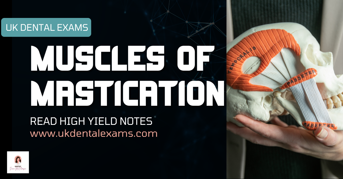
Muscles of Mastication – Origins, Insertions, Innervation & Actions
These four muscles control mandibular elevation, depression, protrusion, retrusion, and lateral movements.
Understanding each muscle’s **origin**, **insertion**, and **action** is critical for TMJ, occlusion, and anesthesia questions in ORE / LDS exams.
| Muscle | Origin | Insertion | Innervation (Branch) | Main Action(s) |
|---|---|---|---|---|
| Temporalis | Triangular muscle with broad attachment to the floor of temporal fossa and deep surface of temporal fascia | Narrow attachment to tip and medial surface of coronoid process and anterior border of ramus of mandible | Deep temporal branches of mandibular nerve (CN V₃) |
Elevates mandible (closing jaws)
Posterior horizontal fibers are the primary retractors of the mandible |
| Masseter | Quadrate muscle attaching to inferior border and medial surface of zygomatic arch & maxillary process of zygomatic bone | Angle and lateral surface of ramus of mandible | Masseteric nerve (branch of CN V₃) |
Elevates mandible; closes jaws
Superficial fibers make limited contribution to protrusion |
| Lateral Pterygoid |
Two-headed muscle from:
(1) Infratemporal surface & crest of greater wing of sphenoid (2) Lateral surface of lateral pterygoid plate |
Superior head: TMJ capsule & articular disc
Inferior head: pterygoid fovea on neck of condyloid process | Lateral pterygoid nerve (from CN V₃) |
Acting bilaterally: protrudes mandible & depresses chin
Acting unilaterally: swings jaw toward contralateral side Alternate unilateral activity → large lateral chewing movements |
| Medial Pterygoid |
Quadrangular two-headed muscle from:
(1) Medial surface of lateral pterygoid plate & pyramidal process of palatine bone (2) Tuberosity of maxilla | Medial surface of ramus of mandible, inferior to mandibular foramen (mirror image of masseter) | Medial pterygoid nerve (from CN V₃) |
Elevates mandible (synergist of masseter)
Contributes to protrusion Alternate unilateral activity → small grinding movements |
**Innervation of Temporalis, Masseter and Lateral Pterygoid: Anterior trunk of Mandibular Nerve (CN V3)
| Muscle | Origin | Fibres | Insertion |
|---|---|---|---|
| Masseter Quadrilateral, covers lateral surface of ramus of mandible, has three layers |
|
|
|
| Temporalis Fan-shaped, fills the temporal fossa |
| Anterior fibers run vertically, middle obliquely, and posterior horizontally. All converge and pass through gap deep to zygomatic arch |
|
| Lateral pterygoid Short, conical, has upper and lower heads |
| Fibres run backwards and laterally and converge for insertion |
|
| Medial pterygoid Quadrilateral, has a small superficial and a large deep head |
| Fibres run downwards, backwards, and laterally. The two heads embrace part of the lower head of lateral pterygoid | Roughened area on the medial surface of angle and adjoining ramus of mandible, below and behind the mandibular foramen and mylohyoid groove |


| Movement | Muscles |
|---|---|
| Depression |
|
| Elevation |
|
| Protrusion (Protraction) |
|
| Retraction | Posterior fibres of temporalis |
| Side-to-side (chewing) movement | Medial and lateral pterygoids of one side contracting alternatively with that of opposite side |


| Movements of Mandible | Muscle(s) |
|---|---|
| Elevation (close mouth) | Temporalis, masseter, and medial pterygoid |
| Depression (open mouth) | Lateral pterygoid, suprahyoid, and infrahyoid muscles* |
| Protrusion (protrude chin) | Lateral pterygoid, masseter, and medial pterygoid* |
| Retrusion (retrude chin) | Temporalis (posterior oblique and near horizontal fibers) and masseter |
| Lateral movements (grinding and chewing) | Temporalis of same side, pterygoids of opposite side, and masseter |
*The prime mover is normally gravity; these muscles are mainly active against resistance.
The lateral pterygoid is the prime mover here, with minor secondary roles played by the masseter and medial pterygoid.

Leave a Reply